Oral, Dental, and Maxillofacial Radiology is a subspecialty field of dentistry that aims to make accurate diagnoses of diseases and plan appropriate treatments.
Oral Diagnosis and Radiology is the first specialized field patients encounter when they visit a dentist with complaints about oral, dental, or soft tissue issues. Managing the process from meeting the patient to treatment planning with accurate information is important for breaking the "dentist" phobia and gaining the trust of patients in every way.
Determining the medical issues in the patient's past and present history, conducting a detailed intraoral and extraoral examination, interpreting periapical-panoramic-tomographic X-ray findings, making accurate differential diagnoses of diseases, and planning the appropriate treatment based on all this data are the skills of this subspecialty field.
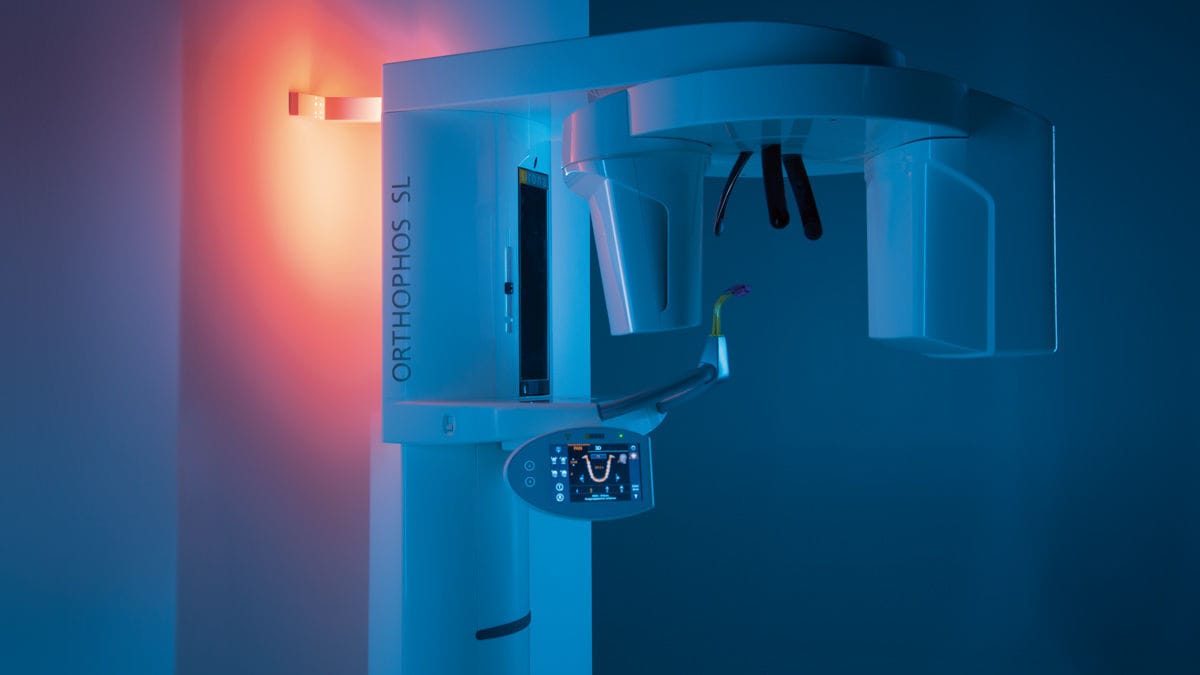
ORTHOPHOS SL 2D
We provide services with X-ray devices that facilitate advanced diagnostic options for endodontics, periodontics, implantology, orthodontics, surgery, and other specialized departments.
Thanks to Sharp Layer Technology (SL), high-resolution panoramic images are not only obtained in the sharp layer, but also enable analysis in the lingual/buccal plane in special cases.
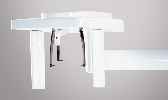
This imaging device has a viewing area starting at 5cm x 5cm and extending up to 11cm x 10cm for upper respiratory tract analysis.
ORTHOPHOS SL 2D, with DCS (Direct Conversion Sensor) sensor and SL technology, provides images that satisfy even dentists with high demands for panoramic imaging. With its 3-point head immobilization, balanced arms, and patented occlusal bite rod, it guarantees unique resolution in every image. Together with the pioneering SIDEXIS 4 software, it offers a unique variety of innovative solutions for clinical workflow.
Our hospital's ORTHOPHOS SL device, which perfectly fits the pleasant atmosphere, provides relaxing ambient lighting with more than 30 colour options, which also has a special feature for patients with claustrophobia, enabling our patients to have a more comfortable, smooth, and fast imaging.
Most of the patients who visit our hospital are hesitant to visit the radiology department due to the amount of radiation from dental X-rays. However, there are some differences between our hospital's ORTHOPHOS SL device and the X-ray devices used in other hospital institutions or clinics:
- By taking quick shots within 15 seconds, the radiation dose the patient receives is cut in half.
- With its sharp X-ray feature, it automatically gathers and combines sharp layers in films according to the patient's personal anatomical structure, providing HD images to our dentists, making it easier for them to make more accurate diagnoses.
- Thanks to the Sharp Layer Technology (SL) technology, high-resolution panoramic images are not only obtained in the sharp layer, but also enable interactive analysis in the lingual/buccal plane in special cases.
- Our X-ray device with HD mode capability allows us to see the thin root canals in the patient's jaw structure with high-quality images even in panoramic films.
- Did you know that the radiation dose from our SIRONA ORTHOPHOS SL 2D/3D X-ray device is less than the radiation dose from X-ray shots taken with other devices used in hospitals and clinics, as well as the radiation dose you receive in your daily life?
In the table below, the radiation doses mentioned are compared with the radiation dose given by our ORTHOPHOS SL device. As a result, it is concluded that each device does not give the same radiation dose, and that the devices used change the amount of radiation received.
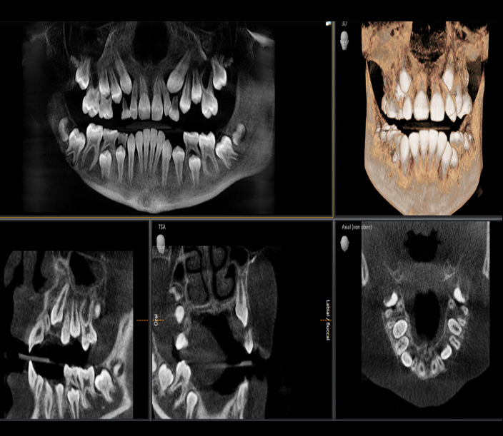
ÜÇÜNCÜ BOYUT (3D) HASTA TEŞHİSLERİNDE FARK YARATIR
Düşük Doz: 2D Görüntü Dozlama aralığında CBCT 3D GÖRÜNTÜLEME
- 2D panoramik x ışınlarının doz düzeyinde 3d görüntüleme sistemine sahip olup hastalarımıza en düşük doz ile ağız içi tomografi görüntüleri elde ediyoruz.
- Düşük Doz modu ile artık 2D X-ışını görüntüleriyle aynı doz aralığında 3D görüntüleri kullanarak endikasyona dayalı tanı artık mümkündür. Optimize edilmiş ön filtreleme, kemik gibi yoğun yapıları çok düşük bir dozda bile korur, böylece ortodonti veya implantoloji gibi birçok uzmanlık alanında kolay ve verimli bir şekilde kullanılabilir.
- Daha kesin tanılar, net açıklamalar: 3D görüntülemenin bir çok avantajı vardır.Üstüste binmiş dişler,umulmadık sinir kanal geçişleri,gizli kökler,gömülü diş veya temporomandibular eklem vakalarında 3D görüntüleme özelliği ile çok sayıda paha biçilmez değerde tanı olanağı sağlamaktayız.
- 11cm x10cm’e kadar tüm çeneyi kapsayarak 20 sn içerisinde hızlı ve en düşük dozda 3D görüntüsü elde etme özelliğine sahip röntgen cihazı ile hastalarımıza yüksek kaliteli hizmet sunuyoruz.
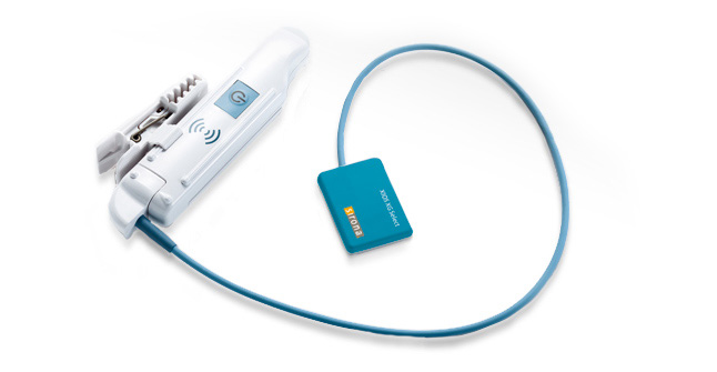
XIOS XG Supreme (PERİAPİKAL RÖNTGEN)
*Endodonti hekimlerin vazgeçilmez görüntüleme cihazı periapikal röntgen cihazları, kanal tedavilerinde, kök uçlarında belirlemeyen lezyon vb gibi durumlarda diş ve diş çevrelerinde oluşan vakaları daha yakından görmeyi kolaylaştıran röntgen cihazlardır.
Aynı zamanda bir kablo sınırları olmadan, opsiyonel WiFi
modülü ile kliniğin her yerinde hastaların teşhis için gereken hareket özgürlüğü sağlayarak kablosuz görüntü aktarımı ile olağanüstü kalitesi ve kolay kullanımlı iş akışı ile donatılmıştır.
*Tek tuş hızlı çekimi ile hastanın film görüntüsü hızlıca ekrana düşme özelliğine sahiptir.
Cephalometric (Sefalometrik Röntgen)
- Ortodonti bölümü yani diş teli tedavilerinde alınan filmlerdir.
- Tüm ortodontik ihtiyaçlarını kapsayan geniş ceph (sefalometrik)programları özelliğine sahiptir.
- Genellikle ortodonti bölümüne gelen çocuk hastaları olduğundan çocuk modu ile çekim yapma özelliği ile 2D ve 3D modunda alınan dozun daha düşük seviyesinde görüntü alma özelliğine sahiptir. Bu yüzden çocuk hastalarımıza rahatça film alma şansımızı kolaylaştırıyor.
- 15 sn hızlı çekim özelliğine sahip röntgen cihazıdır.
- 1-2 sn içerisinde hızlı görüntü aktarımına sahiptir.
- Çocuk hastalarımızı daha rahatlatmak amaçlı 30’dan fazla ambiyans ışıklandırma sistemine sahiptir.